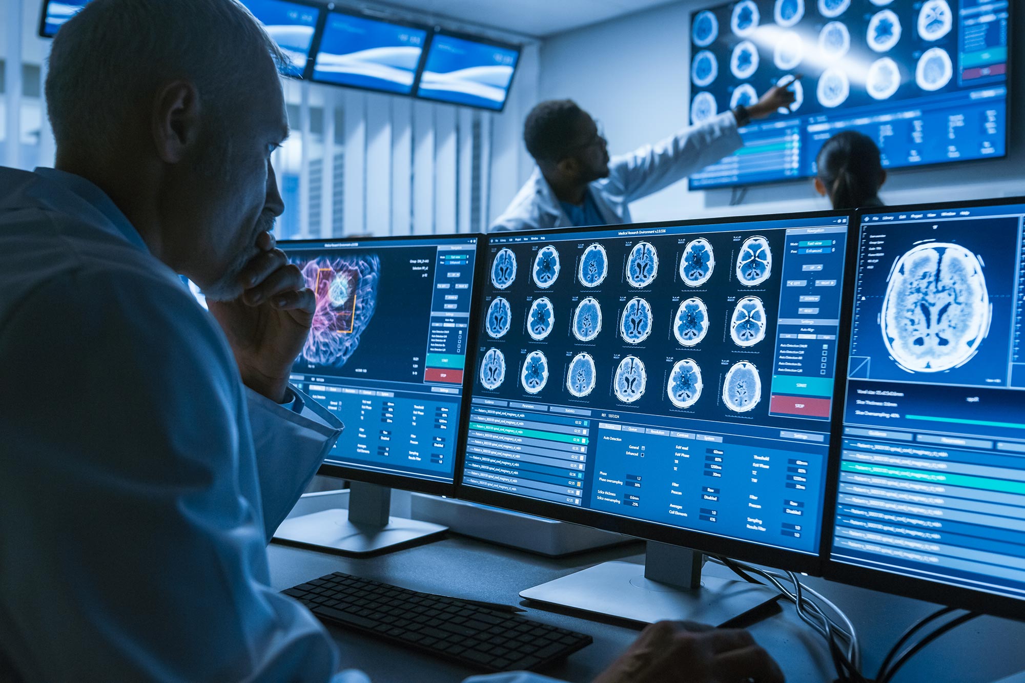
For the important thing time, magnetic resonance imaging shows in vivo brain inflammation. The describe above is an artist’s theory of scientists studying a brain scan.
Utilizing diffusion-weighted magnetic resonance, researchers captured photography of the activation of microglia and astrocytes, two forms of cells inquisitive about neuroinflammation.
The labs of Dr. Silvia de Santis and Dr. Santiago Canals from the Institute of Neurosciences UMH-CSIC (Alicante, Spain) possess faded diffusion-weighted magnetic resonance imaging to describe brain inflammation now no longer only for the important thing time but moreover in huge detail.
Records gathering sequences and surely educated mathematical items are critical to invent this in-depth “X-ray” of inflammation, which would perchance’t be completed with a faded MRI. After growing the methodology, the researchers had been in a position to measure the adjustments in the morphology of the a colossal selection of cell populations contributing to the inflammatory route of in the brain.
This critical discovery, which modified into as soon as recently published in the journal Science Advances and would perchance be a must-want to altering the trajectory of be taught and treatment of neurodegenerative ailments, modified into as soon as made imaginable by an progressive strategy created by the researchers.
Researchers from the UMH-CSIC Neurosciences Institute possess created a original system that allowed them to visualize microglial and astrocyte activation. Credit: IN-CSIC-UMH
The peep, whose first author is Raquel Garcia-Hernández, shows that diffusion-weighted MRI can noninvasively and differentially detect the activation of microglia and astrocytes, two forms of brain cells that are at the source of neuroinflammation and its pattern.
Degenerative brain stipulations including Parkinson’s, quite a bit of sclerosis, Alzheimer’s, and other dementias are serious and traumatic factors to resolve. Considered one of many causes of neurodegeneration and a trust its development is chronic inflammation in the brain, which is attributable to the sustained activation of two forms of brain cells, microglia and astrocytes.
Nonetheless, there is a scarcity of non-invasive approaches in a position to particularly characterizing brain inflammation in vivo. The fresh gold approved is positron emission tomography (PET), on the opposite hand it is complex to generalize and is linked with exposure to ionizing radiation, so its expend is proscribed in susceptible populations and in longitudinal stories, which require the expend of PET many cases over a duration of years, as is the case in neurodegenerative ailments.
Another drawback of PET is its low spatial resolution, which makes it spoiled for imaging cramped structures, with the added drawback that inflammation-particular radiotracers are expressed in quite a bit of cell forms (microglia, astrocytes, and endothelium), making it now no longer doable to distinguish between them.
Within the face of these drawbacks, diffusion-weighted MRI has the peculiar skill to describe brain microstructure in vivo noninvasively and with high resolution by taking pictures the random circulate of water molecules in the brain parenchyma to generate distinction in MRI photography.
Utilizing an progressive strategyIn this peep, researchers from the UMH-CSIC Neurosciences Institute possess developed an progressive strategy that allows imaging of microglial and astrocyte activation in the gray matter of the brain using diffusion-weighted magnetic resonance imaging (dw-MRI).
“Right here’s the important thing time it has been shown that the signal from this form of MRI (dw-MRI) can detect microglial and astrocyte activation, with particular footprints for every cell population. This strategy we’ve faded reflects the morphological adjustments validated post-mortem by quantitative immunohistochemistry,” the researchers reward.
They possess got moreover shown that this methodology is sensitive and particular for detecting inflammation with and without neurodegeneration in explain that both stipulations would possibly also be differentiated. As well, it makes it imaginable to discriminate between inflammation and demyelination characteristics of quite a bit of sclerosis.
This work has moreover been in a position to prove the translational fee of the advance faded in a cohort of healthy humans at high resolution, “wherein we performed a reproducibility diagnosis. The critical association with known microglia density patterns in the human brain supports the usefulness of the model for producing legit glia biomarkers. We contemplate that characterizing, using this methodology, relevant parts of tissue microstructure all the draw in which thru inflammation, noninvasively and longitudinally, can possess an limitless affect on our knowing of the pathophysiology of many brain stipulations and would possibly well modified into fresh diagnostic be conscious and treatment monitoring ideas for neurodegenerative ailments,” highlights Silvia de Santis.
To validate the model, the researchers possess faded a longtime paradigm of inflammation in rats in accordance with intracerebral administration of lipopolysaccharide (LPS). On this paradigm, neuronal viability and morphology are preserved, whereas inducing, first, activation of microglia (the brain’s immune plan cells), and in a delayed system, an astrocyte response. This temporal sequence of cell events allows glial responses to be transiently dissociated from neuronal degeneration and the signature of reactive microglia investigated independently of astrogliosis.
To isolate the impress of astrocyte activation, the researchers repeated the experiment by pretreating the animals with an inhibitor that speedy ablates about 90% of microglia. Therefore using a longtime paradigm of neuronal hurt, they tested whether or now no longer the model modified into as soon as in a position to solve neuroinflammatory “footprints” with and without concomitant neurodegeneration. “Right here’s serious to prove the utility of our advance as a platform for the invention of biomarkers of inflammatory put of residing in neurodegenerative ailments, where both glia activation and neuronal hurt are key players,” they account for.
At closing, the researchers faded a longtime paradigm of demyelination, in accordance with focal administration of lysolecithin, to prove that the biomarkers developed enact now no longer think the tissue alterations customarily stumbled on in brain complications.
Reference: “Mapping microglia and astrocyte activation in vivo using diffusion MRI” by Raquel Garcia-Hernandez, Antonio Cerdán Cerdá, Alejandro Trouve Carpena, Impress Drakesmith, Kristin Koller, Derek Ample. Jones, Santiago Canals and Silvia De Santis, 27 Could perchance perchance perchance neutral 2022, Science Advances.
DOI: 10.1126/sciadv.abq2923

